

Precision-guided surgery using 3D imaging to target a specific point in the brain with minimal disruption.
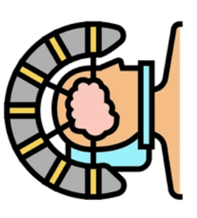
What is stereotactic neurosurgery?
- Stereotactic neurosurgery is an advanced technique used to perform highly precise procedures within the brain
- It combines specialised imaging technology with computer-guided navigation to accurately target specific areas while protecting nearby healthy tissue
- Unlike conventional open surgery, many stereotactic procedures are minimally invasive and may involve smaller cuts (incisions), less discomfort and faster recovery
What are the conditions that may require stereotactic neurosurgery?
Stereotactic neurosurgery may be used to diagnose or treat several conditions affecting the brain and nervous system.
It is commonly performed for:
- Taking a small tissue sample from the brain (biopsy) to identify abnormalities
- Treating abnormal growths within the brain
- Draining infected fluid collections or fluid-filled sacs (cysts) in the brain
- Relieving pressure caused by excess fluid within the brain
- Treating certain types of bleeding (haemorrhages) within the brain
- Evaluating and treating epilepsy or repeated seizures
- Managing movement disorders such as Parkinson’s disease, tremors and involuntary movements
- Treating long-term severe facial pain or long term pain conditions
- Delivering targeted radiation treatment for abnormal blood vessels or selected brain conditions
Why is stereotactic neurosurgery performed?
Doctors may recommend stereotactic neurosurgery when traditional surgery may be risky or when a small abnormal growth (lesion) is located deep within the brain. The technique allows surgeons to reach the target with exceptional precision.
Preparing for surgery
Before surgery, your healthcare team may advise:
- Blood tests
- ECG or chest X-ray if needed
- Review of existing medications
- Fasting for several hours before surgery
- Informing the doctor about allergies or medical conditions
Procedure overview
Step-by-step procedure
Step 1: Placement of the head frame
A specialised frame is gently attached to the patient’s head after the scalp is numbed with local anaesthesia. This frame helps the surgeon accurately identify the treatment area.
Step 2: Brain imaging
Magnetic resonance imaging (MRI) or computed tomography (CT) scans are performed to create detailed images of the brain. These images help the surgical team locate the exact area requiring treatment.
Step 3: Procedure planning
Using the scan images, the neurosurgeon carefully plans the safest and most precise path to reach the targeted area.
Step 4: Preparing for surgery
The patient is positioned comfortably in the operating room. A small area of the scalp is numbed, and a small opening may be made in the skull to access the brain.
Step 5: Targeting the treatment area
Specialised instruments are guided towards the exact treatment area using the stereotactic navigation system.
Step 6: Brain activity monitoring
In some procedures, very thin electrodes may be used to monitor brain activity and confirm the correct target area.
Step 7: Treatment
Depending on the condition being treated, the surgeon may remove a small abnormal area, place an electrode for deep brain stimulation or perform another targeted treatment.
Step 8: Monitoring and recovery
The patient’s condition and vital signs are continuously monitored throughout the procedure to ensure safety and support recovery.
How long does the procedure take?
The duration of stereotactic neurosurgery depends on the type and complexity of the procedure. Most procedures usually take between one and four hours. Some complex cases may take longer.
Benefits of stereotactic neurosurgery
- Minimally invasive procedure: Stereotactic neurosurgery usually requires only small openings or cuts, which may help reduce pain and support quicker recovery
- High precision: Advanced imaging and computer-guided navigation help surgeons accurately target the affected area while protecting nearby healthy tissue
- Lower risk of complications: The minimally invasive approach may reduce the risk of bleeding, infection and damage to surrounding healthy part of the brain
- Personalised treatment: The technique can be adapted for different neurological conditions and individual patient needs
Risks of stereotactic neurosurgery
Although stereotactic neurosurgery is generally considered safe, complications may occasionally occur.Possible side effects after the procedure include:
- Tiredness
- Headache or nausea
- Temporary swelling in the brain
- Mild discomfort at frame sites
- SeizuresBottom of Form

Myths & Facts
Stereotactic neurosurgery always involves major open brain surgery.
Many stereotactic procedures are minimally invasive and may require only small openings or incisions.
The procedure is not safe because it involves the brain.
Stereotactic neurosurgery uses advanced imaging and computer-guided navigation to improve accuracy and reduce damage to nearby healthy brain areas.
All patients undergoing stereotactic neurosurgery need long hospital stays.
Many patients recover quickly and may return home within a short period, depending on the procedure performed.
Stereotactic neurosurgery is only used for brain tumours.
It may also be used for epilepsy, Parkinson’s disease, tremors, long- term painful conditions and other neurological conditions.
Frequently Asked Questions
Is the procedure painful or uncomfortable?
Most patients experience minimal discomfort during stereotactic neurosurgery because local anaesthesia, sedation or general anaesthesia may be used during the procedure. Pain after the procedure is usually less compared to traditional open surgery.
Will I be awake during the procedure?
Some stereotactic procedures are performed while the patient is awake with local anaesthesia, while others may require general anaesthesia. Your doctor will explain the best option for you.
How long does the treatment take?
The duration of the procedure can vary depending on the complexity of the case, but most stereotactic surgeries take between one to four hours.
What are the potential side effects?
Side effects after stereotactic neurosurgery are usually mild and temporary. Some patients may experience headache, tiredness, mild swelling or temporary neurological symptoms after the procedure. Serious complications are uncommon.
Will I need follow-up visits after the procedure?
Yes. Follow-up visits and imaging scans may be needed to monitor recovery and treatment results.
Speak to a neurology specialist
Get an expert opinion and a personalised treatment plan at Sterling Hospitals.


